

Last Updated: September 11, 2026
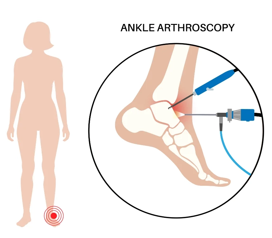
Wrist and ankle arthroscopy is a minimally invasive surgical technique that allows orthopedic surgeons to evaluate and treat certain cartilage, ligament, and soft-tissue problems through small incisions. Fellowship-trained extremity surgeons in Los Angeles use a tiny camera and narrow instruments to work through a few small cuts instead of one large incision. This approach treats conditions such as a wrist TFCC tear or an ankle osteochondral lesion of the talus through small incisions, with most procedures performed on an outpatient basis. LA Orthopedics pairs this precise, minimally invasive technique with a clear recovery plan built around each patient’s specific injury.
Wrist clicking during a twist, an ankle that catches mid-stride, and pain that ignores rest and bracing are signs worth checking out, not living with. This guide covers how wrist and ankle arthroscopy in Los Angeles works, what conditions it treats, and what recovery actually looks like week by week.
If persistent wrist or ankle symptoms are limiting your activity, an orthopedic evaluation can help determine the cause and whether arthroscopy is an appropriate treatment option.
Understanding Small-Joint Arthroscopy: Wrist & Ankle Anatomy
The wrist and ankle are among the most-worked joints in the body. Each uses a dense group of small bones, ligaments, and cartilage to bear heavy loads while still allowing a wide range of motion. The space inside these joints is small, so even a small tear or loose piece of cartilage can cause pain, clicking, or a feeling that the joint might give way.
Because these joints contain many small structures within a limited space, arthroscopy can provide direct visualization through small surgical portals. Arthroscopy provides a direct view of structures inside the joint and can sometimes identify abnormalities that are difficult to fully characterize with imaging alone.
Radiocarpal Joint Anatomy & Triangular Fibrocartilage Complex (TFCC)
The radiocarpal joint is the main wrist joint, where the end of the radius meets several of the small carpal bones of the wrist. The scapholunate ligament is the key ligament that holds the two wrist bones in alignment as you grip and twist. On the pinky side is the triangular fibrocartilage complex, a cushion that absorbs shock between the bones of the forearm and hand.
This cushion, the TFCC, bears a lot of the load on the wrist when you grip and push. It can rip from a fall or years of repeated twisting. If it does, the wrist loses its shock-absorbing ability and often hurts or clicks, such as when you turn a doorknob.
Talocrural Joint Anatomy & Ankle Cartilage Architecture
The joint that bends when you flex your foot up and down is known as the talocrural joint. The two bones of the lower leg sit on top of the talus bone. The talus is covered with a smooth layer of cartilage, which allows the ankle to bend without friction. Another reason this damage heals slowly is the cartilage’s small blood supply.
The outer front part of the talus is often damaged in a rolled ankle sprain, while the inner side slowly wears away from repeated stress. If the cartilage in either area is damaged, the smooth surface becomes rough, leading to catching, swelling, and a deep ache when you bear weight on the ankle.
Advantages of Small-Joint Arthroscopy Over Open Surgery
Surgeons use special cameras called arthroscopes that are only 1.9mm to 2.7mm wide so they can work through cuts just larger than a pencil tip. The small portals are designed to minimize disruption to surrounding tissues compared with larger open incisions.
- Smaller portals may reduce soft-tissue disruption compared with larger open incisions
- Smaller incisions generally result in smaller scars
- Direct visualization allows the surgeon to assess structures inside the joint
- Depending on the procedure, a minimally invasive approach may allow earlier rehabilitation
Primary Indications for Wrist Arthroscopy
Wrist arthroscopy may be considered for selected patients whose symptoms persist despite appropriate nonsurgical treatment and whose examination or imaging suggests a problem that may benefit from arthroscopic evaluation or treatment.
Triangular Fibrocartilage Complex (TFCC) Tears & Debridement
A TFCC tear can cause pain on the pinky side of the wrist, clicking or catching with rotation, and discomfort during gripping or weight-bearing through the hand. During surgery, the surgeon checks the tissue closely, since some frayed edges only need smoothing while other true tears need a full repair.
For frayed tissue, the surgeon trims away the damaged part, a process called debridement. For a fresh tear with healthy tissue nearby, the surgeon may instead repair the torn tissue, depending on the location and pattern of the tear.
Scapholunate Ligament Tears & Wrist Instability
This ligament connects two small wrist bones. It stabilizes the wrist during heavy use (lifting, pushing, etc.). A tear can be a small one or a full rupture. The treatment depends on the severity and chronicity of the injury. Selected injuries may be treated arthroscopically, sometimes with temporary fixation, while more advanced instability may require ligament repair or reconstruction.
Selected acute or less advanced injuries may be treated with arthroscopic assessment and temporary fixation, depending on the tear pattern and degree of instability.
Ganglion Cyst Resection & Arthroscopic Synovectomy
Ganglion cysts are small fluid-filled sacs that tend to develop on the back of the wrist from a stalk that goes back to the joint. They can be a bump or remain silent and painful, with no visible lump. An arthroscopic excision both identifies the cyst’s origin and removes the stalk to minimize recurrence.
Arthroscopic synovectomy treats ongoing swelling of the joint lining, which can follow an injury or a condition like rheumatoid arthritis. Removing inflamed synovial tissue may help reduce symptoms in appropriately selected patients while limiting disruption to surrounding structures.
Primary Indications for Ankle Arthroscopy
Ankle arthroscopy surgery in Los Angeles treats a different but equally common set of problems, many starting with a past sprain, repeated impact, or slow cartilage wear. As with the wrist, arthroscopy lets the surgeon see the joint directly and, when appropriate, treat certain abnormalities during the same procedure.
Osteochondral Lesions of the Talus (OLT) & Microfracture
Arthroscopy can treat an osteochondral lesion of the talus, damage to the cartilage, and bone on the talus surface that usually follows a bad ankle sprain or fracture. Osteochondral lesions can occur after ankle sprains or fractures and may contribute to persistent deep ankle pain, swelling, catching, or stiffness after the original injury has healed.
During surgery, the surgeon removes loose cartilage pieces, and curettage clears out any bone cyst underneath. For selected smaller lesions, the surgeon may perform bone marrow stimulation, often called microfracture, to encourage formation of repair tissue. The best treatment depends on the lesion’s size, location, depth, underlying bone, and previous treatment.
Anterior & Posterior Ankle Impingement Syndromes
Ankle impingement happens when bone spurs or thick soft tissue get pinched inside the joint during movement, causing pain at the end of motion. Front, or anterior, impingement, sometimes called footballer’s ankle, comes from repeated forced bending and is common in athletes who cut and pivot. During surgery, the surgeon shaves down the bone spur and clears away thick tissue in the joint.
Back, or posterior, impingement often involves an extra-small bone called the os trigonum, which can cause pain in dancers who often point their toes. When the os trigonum is confirmed as the source of symptoms, arthroscopic removal may reduce impingement and help improve painful motion.
Chronic Ankle Instability & Synovial Debridement
Repeated ankle sprains can also lead to painful scar tissue inside the joint. When this tissue contributes to pain, catching, or restricted motion, it may be removed arthroscopically.
When the ligaments are very loose, surgeons can use arthroscopic tools to tighten the outer ankle structures alongside the debridement. This addresses both looseness and internal scar tissue in a single minimally invasive procedure.
Comparative Decision Framework: Wrist vs. Ankle Arthroscopy
The wrist and ankle differ in size, load, and function, so the arthroscopic approach differs for each. The table below shows how LA Orthopedics distinguishes the two procedures. General examples only; recovery varies significantly depending on the procedure performed.
| Parameter | Wrist Arthroscopy | Ankle Arthroscopy |
|---|---|---|
| Anatomical Target | Radiocarpal & midcarpal joint spaces | Talocrural & subtalar joint spaces |
| Camera Size | Ultra-miniature 1.9mm to 2.4mm arthroscope | Miniature 2.7mm to 4.0mm arthroscope |
| Common Conditions | TFCC tears, scapholunate sprains, ganglion stalks | Talar cartilage lesions (OLT), bone spurs, ankle impingement |
| Portal Locations | Dorsal wrist portals (3-4, 4-5, 6R, 6U) | Anteromedial, anterolateral, and posterior portals |
| Post-Op Weight-Bearing/Use | Varies by procedure; structural repairs generally require longer protection than simple debridement | Varies by procedure; some patients bear weight early, while cartilage or ligament procedures may require longer protection |
| Possible Return-to-Activity Range | Depends on the procedure, healing, rehabilitation progress, and surgeon clearance | Depends on the procedure, healing, rehabilitation progress, and surgeon clearance |
If chronic pain has kept you from work or sport, the foot and ankle surgery team at LA Orthopedics can walk you through which approach fits your case.
What to Expect During Small-Joint Arthroscopy at LA Orthopedics
Patients who choose minimally invasive wrist and ankle surgery in Los Angeles go through a clear set of steps before the procedure, starting with a full exam and a discussion of what the surgeon expects to find.
Outpatient Surgery & Specialized Traction Systems
Wrist and ankle arthroscopy is almost always an outpatient procedure, so you go home the same day. Depending on the procedure and the patient’s needs, anesthesia may involve regional anesthesia, sedation, general anesthesia, or a combination of approaches.
To create working space inside these tight joints, surgeons use traction tools. A finger-trap device gently pulls the wrist apart. An ankle distraction tower does the same job for the ankle. This creates just enough room for the camera and tools to move safely.
Micro-Incisional Portals & High-Definition Video Visualization
The surgeon reaches the joint through small cuts, usually only 2 to 4 millimeters long, placed at exact spots based on the suspected problem. Saline fluid flows through the joint during surgery, keeping the view clear and gently opening the joint space.
A high-definition camera sends live video to a screen, giving the surgeon a zoomed-in view of areas that are hard to see otherwise. Small motorized tools and probes then trim, smooth, or repair tissue, guided by exactly what the camera shows.
Example Arthroscopy Recovery Timeline & Rehabilitation Milestones
Recovery after wrist and ankle arthroscopy isn’t a single countdown to “better”; it moves through distinct stages, each with its own goals for swelling, motion, and strength. How fast a patient moves through them depends largely on what was actually repaired inside the joint.
Recovery varies considerably depending on what the surgeon finds and which procedure is performed. A simple debridement may allow faster progression than cartilage restoration or ligament repair. Your surgeon’s postoperative instructions should take priority over any general timeline.
- Days 1–3: Early recovery often focuses on elevation, swelling control, incision/dressing protection, and prescribed pain management.
- Days 4–7: Depending on the procedure, patients may begin gentle permitted motion and have an early postoperative follow-up.
- Weeks 2–3: The length of immobilization varies considerably by procedure. A simple debridement may require relatively brief protection, while ligament repair, cartilage treatment, or other structural procedures can require a splint, brace, or boot for several weeks. Your surgeon will determine when it is safe to progress.
- Weeks 4–6: Some patients progress range-of-motion and strengthening exercises, while others remain protected longer after ligament or cartilage procedures.
- Months 2–3: Rehabilitation may progress toward strengthening, balance, loading, and activity-specific exercises.
- Months 3–6 and beyond: Return to sports or heavy work depends on healing, strength, function, procedure type, and surgeon clearance.
Why Choose LA Orthopedics for Small-Joint Arthroscopy
The wrist and ankle are unforgiving joints. A surgeon’s skill with this exact anatomy affects both diagnostic accuracy and repair quality. This is why patients look for a small-joint arthroscopy specialist instead of a general orthopedic surgeon.
Fellowship-Trained Hand & Foot/Ankle Specialists
Dr. Shahab Mahboubian leads the extremity team at LA Orthopedics, with fellowship training focused specifically on the wrist, hand, ankle, and foot. This specialty focus shapes every part of patient care, from diagnosis to building a surgical plan around each patient’s anatomy.
Advanced HD Small-Joint Visualization & Biologics
LA Orthopedics uses 4K high-definition cameras and small motorized tools built for the tight spaces of the wrist and ankle, giving a clearer view than older systems allowed.
In selected cases, the surgeon may discuss orthobiologic treatments such as platelet-rich plasma (PRP). Whether these treatments are appropriate depends on the diagnosis and treatment goals, and evidence varies by condition.
Integrated Physical Therapy & Hand Therapy Network
Surgery is only part of a good outcome. LA Orthopedics works with Certified Hand Therapists and physical therapy centers across greater Los Angeles, keeping care lined up with your surgeon’s plan.
Frequently Asked Questions (FAQ)
How long does wrist or ankle arthroscopy surgery take?
The length of wrist or ankle arthroscopy varies depending on what the surgeon needs to evaluate or repair. Some straightforward arthroscopic procedures may take about 30 to 60 minutes, while more involved repairs can take longer. Patients also spend additional time at the surgery center for preparation, anesthesia, and recovery.
How long do I need to wear a splint or boot after arthroscopy?
The length of immobilization varies considerably by procedure. A simple debridement may require relatively brief protection, while ligament repair, cartilage treatment, or other structural procedures can require a splint, brace, or boot for several weeks. Your surgeon will determine when it is safe to reduce or discontinue immobilization based on what was treated and how you are healing.
When can I bear weight after ankle arthroscopy at LA Orthopedics?
Patients who have simple tissue trimming or bone spur removal can often bear weight in a removable boot within 24 to 48 hours. Those who have microfracture may need 4 to 6 weeks of no weight on the foot and may use crutches.
Is wrist or ankle arthroscopy painful during recovery?
Postoperative pain varies with the procedure performed. Your surgical team will provide a pain-management plan that may include elevation, ice when appropriate, medications, and regional anesthesia in selected cases.
What is a TFCC tear, and can it be fixed with wrist arthroscopy?
The triangular fibrocartilage complex is a cushion of cartilage on the pinky side of the wrist. During arthroscopic surgery, a surgeon can trim away painful, frayed edges or stitch torn cartilage back to the bone, depending on the tear pattern.
Will small-joint arthroscopy leave noticeable scars?
Arthroscopy does leave small scars at the portal sites, but they are typically much smaller than an incision used for open surgery. Scar appearance varies by patient and healing.
When can I drive after wrist or ankle arthroscopy?
Driving clearance depends on the extremity involved, immobilization, medications, strength, reaction time, ability to control the vehicle safely, and surgeon instructions.
What happens if I leave a TFCC tear or talar cartilage lesion untreated?
Some untreated injuries can continue to cause pain, catching, or instability and, depending on the type and severity of damage, may contribute to progressive joint degeneration over time. Other injuries remain stable or can be managed without surgery, which is why treatment should be based on the individual diagnosis.
Schedule Your Small-Joint Evaluation at LA Orthopedics
Wrist and ankle arthroscopy has become a reliable option for patients who haven’t found relief with rest, bracing, and physical therapy alone. Small-joint arthroscopy gives surgeons a direct view inside the joint and, when appropriate, the ability to treat certain findings during the same procedure. For appropriately selected patients, arthroscopy can combine direct visualization of the joint with treatment through small incisions. Whether an arthroscopic or open approach is more appropriate depends on the specific injury and procedure required.
Every case is different, and the right treatment plan depends on your specific anatomy, injury pattern, and goals, something only a hands-on evaluation can determine. If chronic wrist or ankle pain, clicking, or weakness has been limiting your daily life or your sport, don’t wait for it to get worse. Schedule a consultation with the fellowship-trained extremity team at LA Orthopedics to learn what’s causing your symptoms and what your options are.
CONTACT LA ORTHOPEDICS
LA Orthopedics
Location: Los Angeles, California
This guide provides general medical information and is not formal medical advice. Individual diagnostic findings and recovery timelines vary. Consult a licensed orthopedic surgeon or extremity specialist for an accurate diagnosis and personalized treatment plan.